Human non pigmented skin slide labeled – Delving into the realm of human non-pigmented skin slide labeled, this comprehensive guide unravels the intricacies of skin anatomy, histopathological techniques, cellular components, and their significance in diagnosing and managing skin conditions. As we embark on this journey, we will uncover the profound role of histopathology in deciphering the health and integrity of our skin.
The subsequent paragraphs will delve into the complexities of skin histology, unraveling the layers of the epidermis, dermis, and hypodermis. We will explore the techniques employed in preparing and staining skin slides, gaining insights into the significance of H&E staining and other methods.
Furthermore, we will investigate the cellular components of non-pigmented skin, examining the functions and interactions of keratinocytes, melanocytes, Langerhans cells, and other skin cells.
Skin Anatomy and Histology

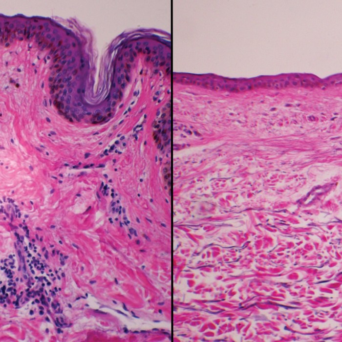
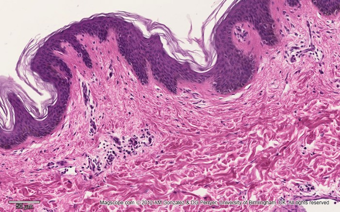
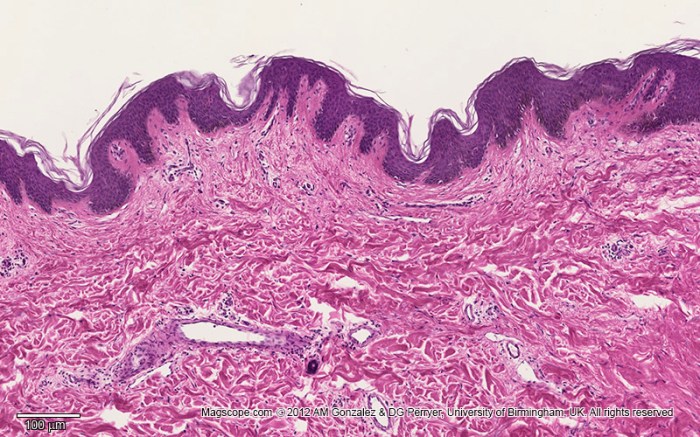
Human non-pigmented skin consists of three distinct layers: the epidermis, dermis, and hypodermis. The epidermis is the outermost layer and is composed of keratinized cells that protect the body from external factors. The dermis lies beneath the epidermis and contains connective tissue, blood vessels, and nerve endings.
The hypodermis is the deepest layer and is composed of fat cells that provide insulation and support.
Epidermis, Human non pigmented skin slide labeled
The epidermis is composed of five layers of cells: the stratum corneum, stratum lucidum, stratum granulosum, stratum spinosum, and stratum basale. The stratum corneum is the outermost layer and is composed of dead cells that are filled with keratin. The stratum lucidum is a thin layer of cells that lies beneath the stratum corneum and is only found in thick skin.
The stratum granulosum is a layer of cells that contains granules of a protein called keratohyalin. The stratum spinosum is a layer of cells that is characterized by its spiny appearance. The stratum basale is the deepest layer of the epidermis and is composed of cuboidal cells that are attached to the basement membrane.
Dermis
The dermis is composed of two layers: the papillary dermis and the reticular dermis. The papillary dermis is the outermost layer and is composed of loose connective tissue. The reticular dermis is the deeper layer and is composed of dense connective tissue.
The dermis contains blood vessels, nerve endings, and hair follicles.
Hypodermis
The hypodermis is composed of fat cells that are arranged in lobules. The hypodermis provides insulation and support to the body.
Histopathological Analysis of Non-Pigmented Skin
Histopathological analysis is the microscopic examination of tissue samples to diagnose diseases. Skin biopsies are commonly used to diagnose skin conditions. Skin biopsies can be obtained from any area of the skin and are typically performed by a dermatologist.
The most common staining method used in histopathology is hematoxylin and eosin (H&E) staining. H&E staining allows for the visualization of the different cellular components of the skin. Other staining methods can be used to highlight specific features of the skin, such as immunohistochemistry and special stains.
Histopathological analysis can be used to diagnose a wide range of skin conditions, including skin cancer, psoriasis, eczema, and acne.
Cellular Components of Non-Pigmented Skin

The cellular components of non-pigmented skin include keratinocytes, melanocytes, Langerhans cells, and fibroblasts.
Keratinocytes are the most common cells in the epidermis. They produce keratin, which is a protein that helps to protect the skin from external factors. Melanocytes are cells that produce melanin, which is a pigment that gives skin its color.
Langerhans cells are immune cells that help to protect the skin from infection. Fibroblasts are cells that produce collagen and elastin, which are proteins that give the skin its strength and elasticity.
Pathophysiology of Skin Conditions
Skin conditions can be caused by a variety of factors, including genetics, environmental factors, and lifestyle choices. Some of the most common skin conditions include psoriasis, eczema, and acne.
Psoriasis is a chronic skin condition that is characterized by red, scaly patches on the skin. Eczema is a chronic skin condition that is characterized by dry, itchy skin. Acne is a common skin condition that is characterized by pimples and blackheads.
The pathophysiology of skin conditions is complex and involves a variety of factors. In psoriasis, the immune system attacks the skin cells, causing them to grow and divide too quickly. In eczema, the skin barrier is damaged, allowing water to escape and irritants to enter.
In acne, the hair follicles become blocked, causing bacteria to grow and produce pus.
Diagnostic Applications of Histopathology: Human Non Pigmented Skin Slide Labeled
Histopathology is a valuable tool for diagnosing skin cancer and other skin diseases. Histopathological analysis can help to differentiate between different types of skin cancer, such as basal cell carcinoma, squamous cell carcinoma, and melanoma. Histopathological analysis can also help to diagnose other skin conditions, such as psoriasis, eczema, and acne.
In some cases, histopathological analysis is the only way to diagnose a skin condition. For example, histopathological analysis is the only way to definitively diagnose melanoma.
Advancements in Skin Histopathology

There have been a number of advancements in skin histopathology in recent years. These advancements have led to improved accuracy and efficiency in the diagnosis of skin diseases.
One of the most significant advancements in skin histopathology is the development of immunohistochemistry. Immunohistochemistry is a technique that uses antibodies to identify specific proteins in the skin. Immunohistochemistry can be used to diagnose a wide range of skin conditions, including skin cancer, psoriasis, and eczema.
Another significant advancement in skin histopathology is the development of digital pathology. Digital pathology involves the use of digital images of skin biopsies to make a diagnosis. Digital pathology allows for the easy sharing of images with other pathologists and for the use of computer-aided diagnosis tools.
Essential Questionnaire
What is the significance of H&E staining in skin histopathology?
H&E staining is a fundamental technique in skin histopathology as it allows for the visualization of cellular structures and components. It provides a clear distinction between the nucleus and cytoplasm, enabling pathologists to identify and characterize different cell types and assess their interactions.
How does histopathological analysis contribute to diagnosing skin cancer?
Histopathological analysis plays a crucial role in diagnosing skin cancer by examining the cellular and structural changes within the skin. It helps differentiate between benign and malignant lesions, determine the type and stage of cancer, and guide appropriate treatment strategies.
What are the limitations of histopathological analysis?
While histopathological analysis is a powerful diagnostic tool, it has certain limitations. It requires a biopsy, which can be invasive and may not always provide a representative sample. Additionally, the interpretation of histopathological findings can be subjective and may vary among pathologists.